
It is a diagnostic procedure that uses two different forms of ultrasonography - Doppler ultrasonography and conventional ultrasonography - to search for carotid artery blockages.
Conventional ultrasonography, also known as B-mode ultrasound, creates an image of your blood vessel anatomy by reflecting sound waves off your blood vessels. Doppler ultrasonography tracks moving objects with the use of sound waves. This makes it possible for your doctor to observe the flow of blood through your blood vessels.
The results of this non-invasive treatment provide important information on the health of the carotid arteries, which feed blood to the head, neck, and brain.
Overview
The Carotid Doppler Ultrasound is a specialized imaging technique that utilizes sound waves to create detailed images of the carotid arteries. These arteries, which run down each side of the neck, are in charge of bringing blood that is high in oxygen from the heart to the brain.
Atherosclerosis, or plaque accumulation, can cause these arteries to constrict, which can result in major health issues like stroke or transient ischemic attack (TIA).
A Carotid Doppler Ultrasound is primarily used to evaluate the blood flow via these arteries and identify any anomalies, such as blockages or narrowing (stenosis). This is a simple and painless procedure. Healthcare providers can assess the risk of stroke and create effective treatment strategies with the use of this method as it provides real-time images and data on blood flow velocity.
Reasons You May Need a Carotid Ultrasound
A Carotid Doppler Ultrasound may be recommended for individuals who are at risk of developing cardiovascular disease or have symptoms suggestive of carotid artery problems.
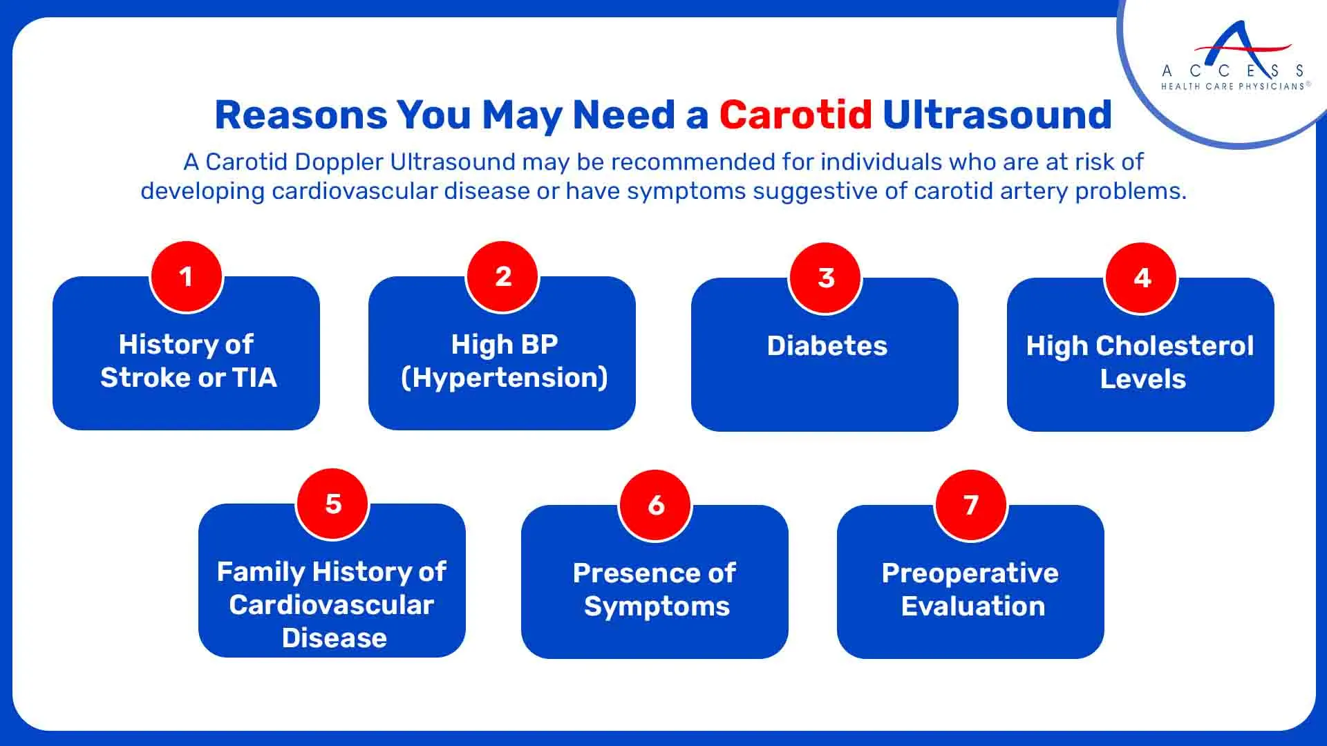
Some common reasons why a healthcare provider may order this test include:
- History of Stroke or TIA:
A carotid Doppler ultrasound can be performed on patients who have had a stroke or transient ischemic attack to evaluate the extent of carotid artery blockage and estimate the risk of subsequent occurrences.
- High Blood Pressure (Hypertension):
Plaque accumulation in the arteries, particularly the carotid arteries, can be accelerated by uncontrolled hypertension (high blood pressure). Assessing the degree of artery damage brought on by high blood pressure can be facilitated by a Doppler ultrasound.
- Diabetes:
Individuals with diabetes are at an increased risk of developing cardiovascular disease. Carotid ultrasound can aid in the early detection of arterial abnormalities in diabetic patients.
- High Cholesterol Levels:
Increased blood cholesterol levels have been linked to the development of arterial plaque. Ultrasound assessment of the carotid arteries can reveal important details regarding the degree and existence of atherosclerosis.
- Family History of Cardiovascular Disease:
Patients with a family history of stroke or heart disease may undergo screening tests such as a Carotid Doppler Ultrasound to evaluate their risk factors and initiate preventive measures if necessary.
- Presence of Symptoms:
A carotid artery issue may be indicated by symptoms such as momentary weakness or numbness on one side of the body, trouble speaking, or abrupt changes in vision. A Doppler ultrasonography is useful in determining the underlying reason in these situations.
- Preoperative Evaluation:
Patients scheduled to undergo certain surgical procedures, particularly those involving the heart or major blood vessels, may require a preoperative assessment of carotid artery health to minimize the risk of perioperative complications.
How Do You Prepare?
Preparing for a Carotid Doppler Ultrasound is generally straightforward and typically does not require any special dietary restrictions or fasting.
However, it is essential to follow any specific instructions provided by your healthcare provider or the imaging facility where the procedure will be performed.
Here are some general guidelines to help you prepare:
- Inform Your Healthcare Provider:
Inform your healthcare provider about all of the medications you use, including prescription, over-the-counter, and dietary supplements, before the ultrasound is scheduled. Before the treatment, it might be necessary to temporarily stop taking some drugs or alter others, including blood thinners.
- Wear Comfortable Clothing:
You will be asked to remove any jewelry, necklaces, or clothing that may interfere with the ultrasound imaging. It is advisable to wear loose-fitting clothing that can be easily removed or adjusted during the examination.
- Follow Dietary Guidelines:
While there are usually no specific dietary restrictions for a Carotid Doppler Ultrasound, it is essential to stay hydrated and maintain your usual eating habits unless instructed otherwise by your healthcare provider.
- Arrive on Time:
Plan to arrive at the imaging facility or hospital at least 15-30 minutes before your scheduled appointment time. This allows ample time for registration and any additional paperwork that may be required.
- Bring Necessary Documents:
Remember to bring your insurance card, identification, and any referral or authorization forms provided by your healthcare provider. Having these documents readily available can streamline the check-in process.
- Communicate Any Concerns:
Do not hesitate to speak with your primary care provider or the imaging technologist doing the ultrasound if you have any questions or concerns regarding the process. They can allay your worries and make sure you're comfortable during the test.
How Does It Work?
A Carotid Doppler Ultrasound employs a technique called Doppler ultrasound to assess blood flow within the carotid arteries and detect any abnormalities.
During the procedure, a specially trained technologist, known as a sonographer, will guide you through the following steps:
- You will be asked to lie on your back on an examination table, with your head tilted slightly backward and turned to one side to expose the side of your neck being examined.
- A clear, water-based gel will be applied to the skin over the area of interest (usually the front of the neck). This gel helps to transmit sound waves and ensures good contact between the ultrasound probe and your skin.
- The sonographer will then gently press a handheld device called a transducer against your skin and move it back and forth over the carotid arteries. The transducer emits high-frequency sound waves that bounce off the blood cells flowing through the arteries.
- As the sound waves travel through the tissues of the neck, they are reflected back to the transducer and converted into real-time images displayed on a monitor. These images show the structure of the carotid arteries, including the presence of plaque, narrowing, or other abnormalities.
- In addition to visualizing the arteries, the Doppler technique allows the sonographer to assess the speed and direction of blood flow. Color-coded images or spectral waveforms may be used to visualize blood flow patterns and detect any areas of turbulence or obstruction.
- The sonographer will capture images and measurements of the carotid arteries at various angles and locations to obtain a comprehensive evaluation of blood flow dynamics and arterial anatomy.
What Happens After a Carotid Ultrasound?
After undergoing a Carotid Doppler Ultrasound, you can expect the following:
- In many cases, the preliminary results of the ultrasound will be available immediately, allowing your healthcare provider to discuss the findings with you during your visit.
- If the ultrasound reveals any abnormalities or areas of concern, your healthcare provider may recommend additional tests or imaging studies to gather more information.
- Depending on the results of the ultrasound and your overall health status, your healthcare provider may schedule a follow-up appointment to discuss the next steps in your care.
In conclusion, the Carotid Doppler Ultrasound is a valuable diagnostic tool for evaluating the health of the carotid arteries and assessing the risk of stroke and other cardiovascular events.
As always, it is essential to consult with your healthcare provider to determine if a Carotid Doppler Ultrasound is appropriate for your specific medical needs and concerns. Connect with Access Health Care Physicians to learn more.
Frequently Asked Questions
Your healthcare provider may recommend a Carotid Doppler Ultrasound if you have symptoms suggestive of carotid artery disease.
No, a Carotid Doppler Ultrasound is a painless and non-invasive procedure. You may feel slight pressure or discomfort as the ultrasound probe is applied to your neck, but it will not cause any significant pain.
The duration of a Carotid Doppler Ultrasound typically ranges from 30 to 60 minutes, depending on the complexity of the examination.

