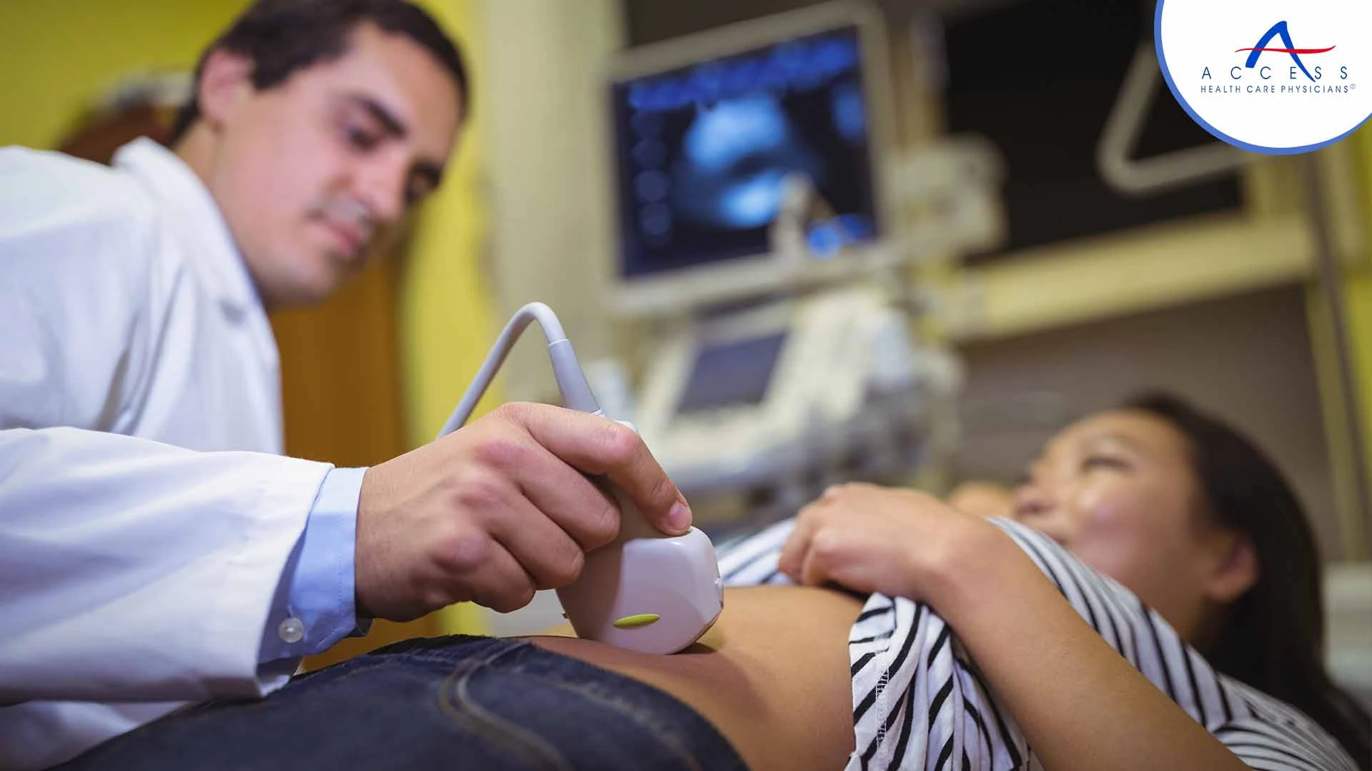
Abdominal ultrasound is a useful diagnostic tool used by medical practitioners to evaluate a range of disorders affecting the organs located within the abdomen. Through the use of sound waves and a non-invasive technique, the abdominal organs can be seen and their shape and function can be better understood.
After applying the ultrasound transducer to the skin, ultrasonic waves pass through the body and into the organs and structures. The transducer will receive the sound waves that bounce off the organs. After the transducer processes the reflected waves, a computer creates an image of the organs or tissues under study. Using ultrasound, blood flow to the abdominal organs can also be evaluated.
Abdominal ultrasonography is a vital component of patient treatment, ranging from determining probable reasons for pain in the abdomen to tracking the advancement of specific illnesses.
Purpose of Abdominal Ultrasound
Abdominal ultrasound serves a multitude of purposes in diagnosing and monitoring conditions affecting the organs within the abdomen.
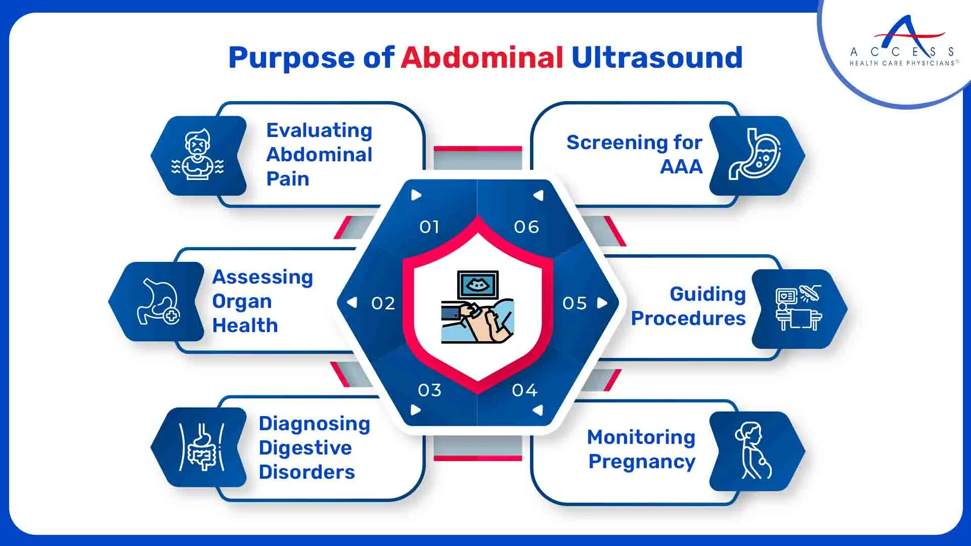
Some common reasons for performing abdominal ultrasound include:
- Evaluating Abdominal Pain:
A common method for determining the source of abdominal pain is abdominal ultrasonography. It assists in locating anomalies that may be causing discomfort in organs like the pancreas, liver, gallbladder, kidneys, and spleen.
- Assessing Organ Health:
Healthcare professionals can evaluate the size, shape, and texture of abdominal organs with this imaging approach. It helps in the detection of anomalies such as inflammation, cysts, and tumors.
- Diagnosing Digestive Disorders:
The diagnosis of several digestive diseases, such as pancreatitis, liver cirrhosis, gallstones, and inflammatory bowel disease, is aided by abdominal ultrasonography.
- Monitoring Pregnancy:
In pregnant women, abdominal ultrasound is used to monitor fetal growth and development, assess the placenta, and detect any abnormalities in the uterus or ovaries.
- Guiding Procedures:
Ultrasound imaging can be used to guide certain procedures such as biopsies, drain placements, and fluid aspirations within the abdomen.
- Screening for Abdominal Aortic Aneurysm (AAA):
Abdominal ultrasound is utilized as a screening tool for detecting AAA, a potentially life-threatening condition characterized by the ballooning of the aorta in the abdomen.
Abdominal Ultrasound Benefits and Risks
While abdominal ultrasound is considered a safe procedure with minimal risks, there are a few considerations to be aware of:
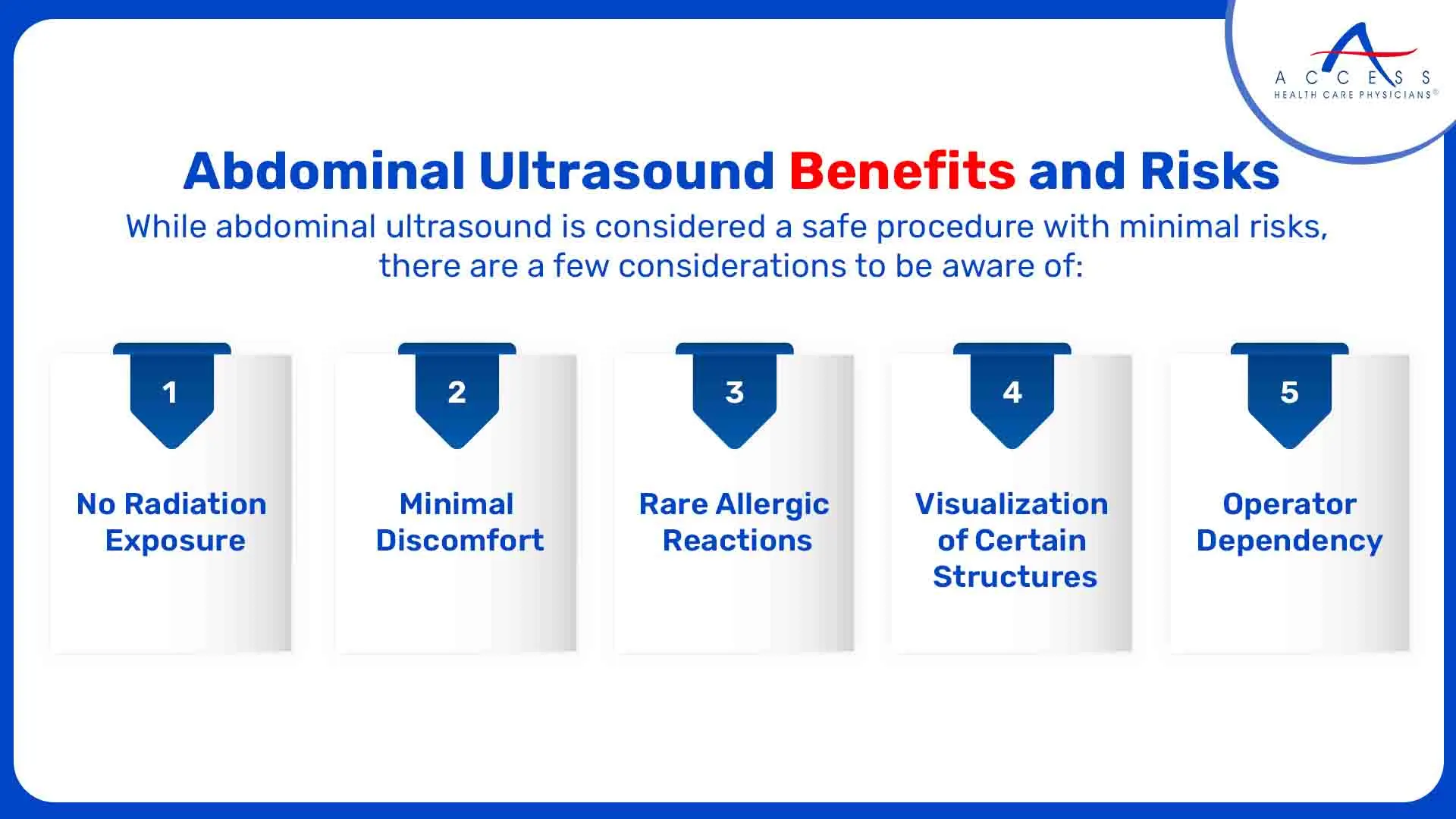
- No Radiation Exposure:
Unlike some imaging techniques such as X-rays or CT scans, abdominal ultrasound does not expose patients to ionizing radiation, making it a safer option, especially for pregnant women and children.
- Minimal Discomfort:
The procedure is generally well-tolerated by patients and does not typically cause discomfort. However, in cases where pressure is applied to enhance imaging, patients may experience mild discomfort.
- Rare Allergic Reactions:
Although rare, some patients may experience allergic reactions to the ultrasound gel used during the procedure. These reactions are usually mild and easily managed.
- Limited Visualization of Certain Structures:
While abdominal ultrasound is effective for visualizing many abdominal organs, it may have limitations in adequately visualizing structures obscured by gas or bones, such as the intestines or pancreas, particularly in obese patients.
- Operator Dependency:
The proficiency and expertise of the ultrasound technologist doing the procedure can have a bearing on the quality of the ultrasound images that are obtained. Untrained operators could find it difficult to capture sharp images, which could cause results to be interpreted incorrectly.
Overall, the benefits of abdominal ultrasound in diagnosing and monitoring abdominal conditions far outweigh the minimal risks associated with the procedure.
Abdominal Ultrasound Preparation
The abdominal ultrasound preparation typically involves minimal effort on the part of the patient. However, following specific guidelines can help ensure the accuracy and effectiveness of the procedure:
- Fasting:
A patient may be told to fast for a specified amount of time before the ultrasound examination, depending on its unique purpose. Abdominal organ visibility is enhanced by fasting, especially the pancreas and gallbladder.
- Medication:
Patients should inform their healthcare provider of any medications they are taking, especially if they are diabetic or taking medication that may affect bowel motility. In some cases, medication adjustments may be necessary before the ultrasound.
- Clothing:
Patients are usually asked to wear comfortable, loose-fitting clothing that can easily be adjusted or removed to expose the abdomen during the procedure.
- Avoiding Gas-Producing Foods:
To minimize interference from gas in the intestines, patients may be advised to avoid gas-producing foods such as beans, broccoli, cabbage, and carbonated beverages in the hours leading up to the ultrasound.
- Arrival Time:
Patients should arrive on time for their appointment and be prepared to provide any necessary information to the healthcare provider or ultrasound technologist.
Abdominal Ultrasound Procedure
The abdominal ultrasound procedure typically follows these general steps:
- Abdominal Ultrasound Preparation:
The patient is positioned lying on their back on an examination table. A water-based gel is applied to the skin over the abdomen to facilitate the transmission of sound waves.
- Transducer Placement:
The ultrasound technologist places a handheld device called a transducer on the abdomen and moves it around to obtain images of the abdominal organs from different angles.
- Image Acquisition:
As the transducer emits high-frequency sound waves, it captures echoes reflected off the internal structures of the abdomen. These echoes are then converted into real-time images displayed on a monitor.
- Evaluation:
The healthcare provider or radiologist interprets the images in real-time, looking for any abnormalities or signs of disease in the abdominal organs.
- Documentation:
The findings of the abdominal ultrasound are documented in a report, which may include measurements of organ size, descriptions of any abnormalities, and recommendations for further evaluation or treatment.
- Completion:
Once the necessary images have been obtained and evaluated, the ultrasound technologist removes the gel from the skin, and the procedure is complete.
The duration of the abdominal ultrasound procedure can vary depending on the specific organs being evaluated and the complexity of the case. In most cases, abdominal ultrasound is a relatively quick and painless procedure, typically lasting between 30 to 60 minutes.
Follow-Up After Abdominal Ultrasound Test
Following an abdominal ultrasound examination, patients may receive further instructions based on the findings and the reason for the test:
- Patients may meet with their healthcare provider or a radiologist to discuss the findings of the ultrasound examination.
- Depending on the results of the abdominal ultrasound, further testing or imaging studies may be recommended to provide additional information or confirm a diagnosis. This could include blood tests, additional ultrasound scans, CT scans, or MRI scans.
- If abnormalities are detected during the ultrasound examination, a treatment plan may be developed in collaboration with the patient's healthcare provider.
- In certain cases, abdominal ultrasound may be used for ongoing monitoring of certain conditions or to assess the effectiveness of treatment over time.
Patients need to follow any recommended follow-up care instructions provided by their healthcare provider to ensure optimal management of their health and well-being.
From assessing abdominal pain to monitoring pregnancy and detecting digestive disorders, abdominal ultrasound plays a crucial role in patient care. With its non-invasive nature, minimal risks, and relatively quick procedure time, abdominal ultrasound provides healthcare providers with valuable insights into the structure and function of abdominal organs.
To learn more about abdominal ultrasound and if you require it, consult with the primary care providers at Access Health Care Physicians.
Frequently Asked Questions
Yes, abdominal ultrasound is considered a safe procedure with minimal risks. Abdominal ultrasound does not expose patients to ionizing radiation, making it a safer option, especially for pregnant women and children.
Patients may be given further advice following the abdominal ultrasound procedure, depending on the results and the purpose of the examination. They can consult with a radiologist or their healthcare practitioner to go over the findings of the ultrasound examination.
Abdominal ultrasound can be used to diagnose and monitor a wide range of conditions such as gallstones, liver cirrhosis, bladder abnormalities, and kidney stones.

